Artificial intelligence digitally stains tissue samples used in pathology, saving labor, time and cost
Tech Xplore | March 05, 2019
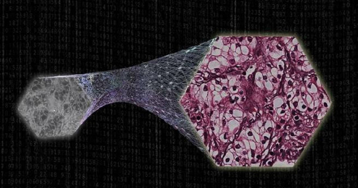
Histopathology is one of the main methods used for diagnosis of disease. Following a medical screening process, a patient can undergo a biopsy, in which a piece of tissue is removed for further inspection and diagnostic analysis. This tissue specimen is then sliced into thin sections that are on the order of a few millionths of a meter in thickness. These thin sections of tissue contain at the microscopic scale the diagnostic information regarding the patient's condition. However, they exhibit almost no contrast under standard light microscopy. To reveal these microscopic features embedded inside tissue and bring visible contrast for inspection by a pathologist, various tissue staining methods have been created in pathology dating back to more than 150 years ago. These tissue staining procedures use different types of colored dyes that specifically label micro-scale structures in tissue, forming color images of specimens, which have been widely used as a gold standard diagnostic method in modern medicine. However, this standard process of staining a tissue specimen is laborious, costly and requires a dedicated laboratory infrastructure, chemical reagents, as well as trained personnel (histotechnologists). Furthermore, currently used staining methods do not preserve tissue samples, which is a limitation since advanced molecular analysis of the tissue sample cannot be easily performed after the initial staining process.